Eye phenotyping is crucial as approximately 40% of donors over the age of 80 with no indication of AMD in referral medical records show some signs of retinal degeneration through post-mortem imaging. Medical records often fail to capture disease stage changes, such as dry converting to wet AMD, which digital pathology can detect.
What MMRI Provides
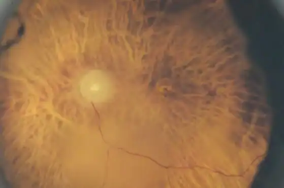
Color fundus photography: 3 magnifications under 2 lighting modalities
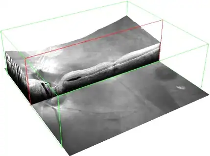
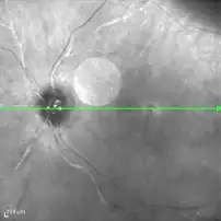
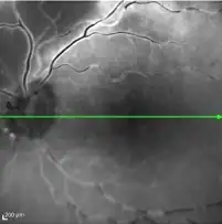
OCT imaging in infrared, red-free, autofluorescence, and volume B-scans or radial scans
Retinal and disease findings
Supports post-mortem phenotyping, including verifying clinical records and identifying cryptic ocular disease
Only available on fixed whole eyes from our On-Demand Sample Library or Perspective Collections